Cartilage Defects
What are Cartilage Defects?
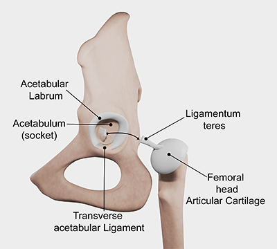
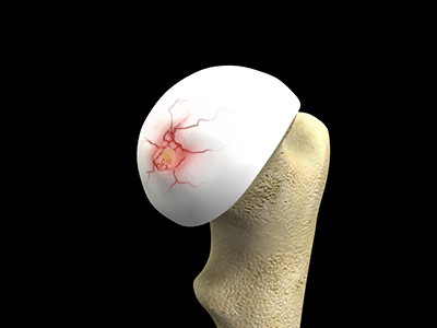
The hip is a ball and socket joint. The bones that make up the hip include the femur (ball) and acetabulum (socket). There is articular cartilage on the end of each of these bones, which is a smooth tissue that allows the bones of the joint to move without causing damage. Damage to this articular cartilage may occur and is referred to as a cartilage defect.
Causes
There are many conditions which can cause cartilage defects in your hip joint:
- Trauma to the hip
- Femoroacetabular impingement syndrome
- Hip dysplasia
- Slipped capital femoral epiphysis
- Avascular necrosis
- Arthritis
Symptoms
A patient who has cartilage defects in their hip joint might experience the following symptoms:
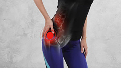
- Significant pain in hip
- Limited range of motion
Diagnosis
As pain in and around your hip can have several causes, diagnosing cartilage defects requires comprehensive testing to be diagnosed confidently and accurately. Your doctor will carefully review your symptoms and medical history and perform a physical examination. Diagnostic tests for this condition include:
- MRI Scan: This study uses a large magnetic field and radio waves to produce images that help in detecting damage to soft tissue, muscles, tendons, and ligaments located in the hip.
- CT Scan: This scan uses multiple X-rays to produce detailed cross-section images of soft tissues, bone, and cartilage structures.
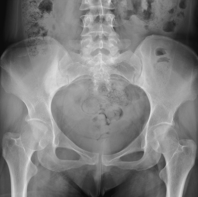
- X-rays: This study uses electromagnetic beams to identify the presence of stress fractures or structural issues.
- Ultrasound: This study uses high-frequency sound waves to produce images of the tissues around the hip.

Treatment Options
Treatment for cartilage defects varies based on age, health, and goals of the patient. Your doctor may initially recommend conservative treatment to help relieve the symptoms.
Examples of conservative treatment include:
- Anti-inflammatory medication: Your doctor may recommend non-steroidal anti-inflammatory drugs (NSAIDs) to reduce pain and inflammation in the tissues.
- Rest and Activity Modification: Reducing or modifying physical activity can reduce the load on the joint and may reduce symptoms
- Ice or Heat: Applying ice or heat can help reduce any swelling or inflammation
- Injections: Your doctor may recommend steroid injections or platelet-rich plasma (PRP) injections to reduce pain and promote healing.
- Physical therapy: After a rest period, exercises are recommended to fix muscular imbalances.
Surgical Options
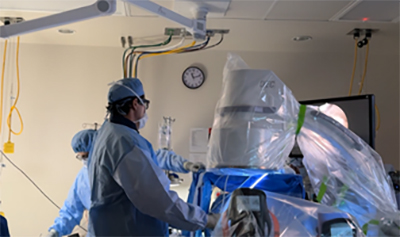
If conservative methods fail to improve a patient's symptoms, surgical treatment may be recommended by an orthopedic provider. Microfracture is an arthroscopic procedure performed to restore injured cartilage. The procedure involves making multiple tiny holes into the bone in the areas where cartilage is damaged. The purpose of making these tiny holes in the bone is to stimulate the patient's bones to regrow the cartilage in that area. Once undergoing a microfracture, a patient can expect to be on crutches for 8 weeks. This period of non-weight bearing allows for orthobiologics to stimulate undisturbed cartilage regrowth.
